Service details
Procedure details and treatment flow
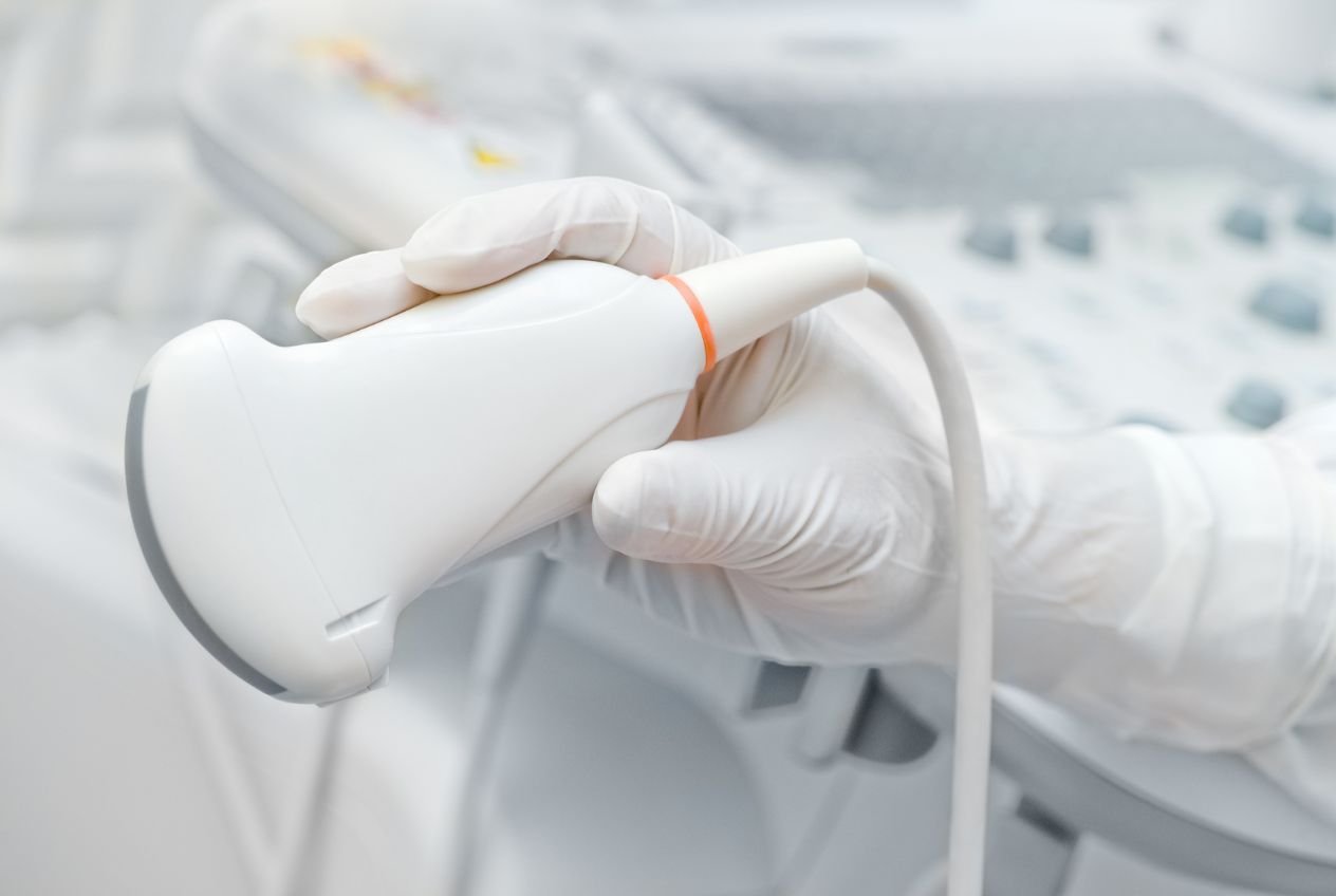
Abdominal and pelvic ultrasound with color doppler examination
Ultrasound of the abdomen and small pelvis is a very useful, painless and completely harmless diagnostic method. Since it uses only high-frequency sound waves to create the image, the patient is not exposed to any X-ray radiation.
In our polyclinic in Kragujevac, this method represents a quick and extremely reliable way for a detailed examination of most structures in the abdominal cavity and pelvis.
It is important to note that air-filled structures (such as the stomach and intestines) are not clearly visible on ultrasound. Endoscopic methods (gastroscopy and colonoscopy) are used for their examination. All other internal organs are shown by ultrasound with great precision.
Mandatory preparation for the examination: In order for the picture of the internal organs to be clear, your cooperation is extremely important.
- Nutrition: The day before the examination, do not eat foods that cause bloating (beans, cabbage, peas, green beans, fresh fruit). Do not eat for at least 4 to 6 hours (preferably 12 hours) before the examination, and avoid coffee because it empties the gallbladder. It is best to do the examination in the morning, on an empty stomach, after emptying the bowels.
- Fluid intake: DO NOT PURINE at least two hours before the examination! When the bladder is full, the bladder itself and all the structures in the pelvis can be seen much better.
* In emergency cases, the examination is done immediately, without any preparation.
What organs and structures are examined?
This examination gives a complete insight into the condition of the solid organs, blood vessels and lymph nodes of the abdominal cavity.
Abdominal and pelvic organs:
- Liver, gallbladder and bile ducts
- Pancreas and spleen
- Kidneys and adrenal glands
- Urinary bladder
- Prostate and seminal vesicles (in men)
- Uterus and ovaries (in women)
- Abdominal wall muscles
Blood vessels and other structures:
- Abdominal aorta
- Portal vein and hepatic veins (in the liver)
- Renal and iliac arteries and veins
- Inferior vena cava and celiac trunk
- Lymph nodes in the abdominal cavity
- The presence of free fluid in the stomach
What can be detected by ultrasound?
Examination is an effective method in detecting and monitoring various diseases. The most common pathological changes that we successfully diagnose are:
- Liver: Fatty liver, hepatitis, cirrhosis (with assessment of portal hypertension by doppler), as well as benign (hemangiomas) and malignant tumors (metastases).
- Bile and Pancreas: Calculus (stone in the gall bladder), inflammation of the gallbladder, acute and chronic pancreatitis, cysts and tumors.
- Kidneys: Stones in the kidney (until fine sand is visible), urinary stasis (hydronephrosis), cysts, tumors, congenital anomalies (descended or horseshoe kidney) and narrowing of the renal arteries.
- Bladder and prostate: Wall thickening, stones, bladder tumors, estimation of prostate size in men.
- Spleen and lymph nodes: Enlargement of the spleen and lymph nodes (most often due to infections or hematological diseases).
- Aorta: Aneurysms (dangerous enlargements) and atherosclerotic plaques (deposits).
- Free liquid: Ascites in the abdominal cavity, purulent collections or effusion of fluid in the lung tissue.
An important note about ultrasound ranges
It is very important to understand that it is only for some of the mentioned diseases, ultrasound is the definitive method (where no additional imaging is required). For a large number of conditions (especially when suspicious tumor changes or metastases are observed), ultrasound represents only an excellent start to the examination. If necessary, our doctor will refer you to additional scans with a scanner (CT, MSCT) or magnetic resonance imaging (MRI) in order to establish an unmistakable and final diagnosis.