Service details
Procedure details and treatment flow
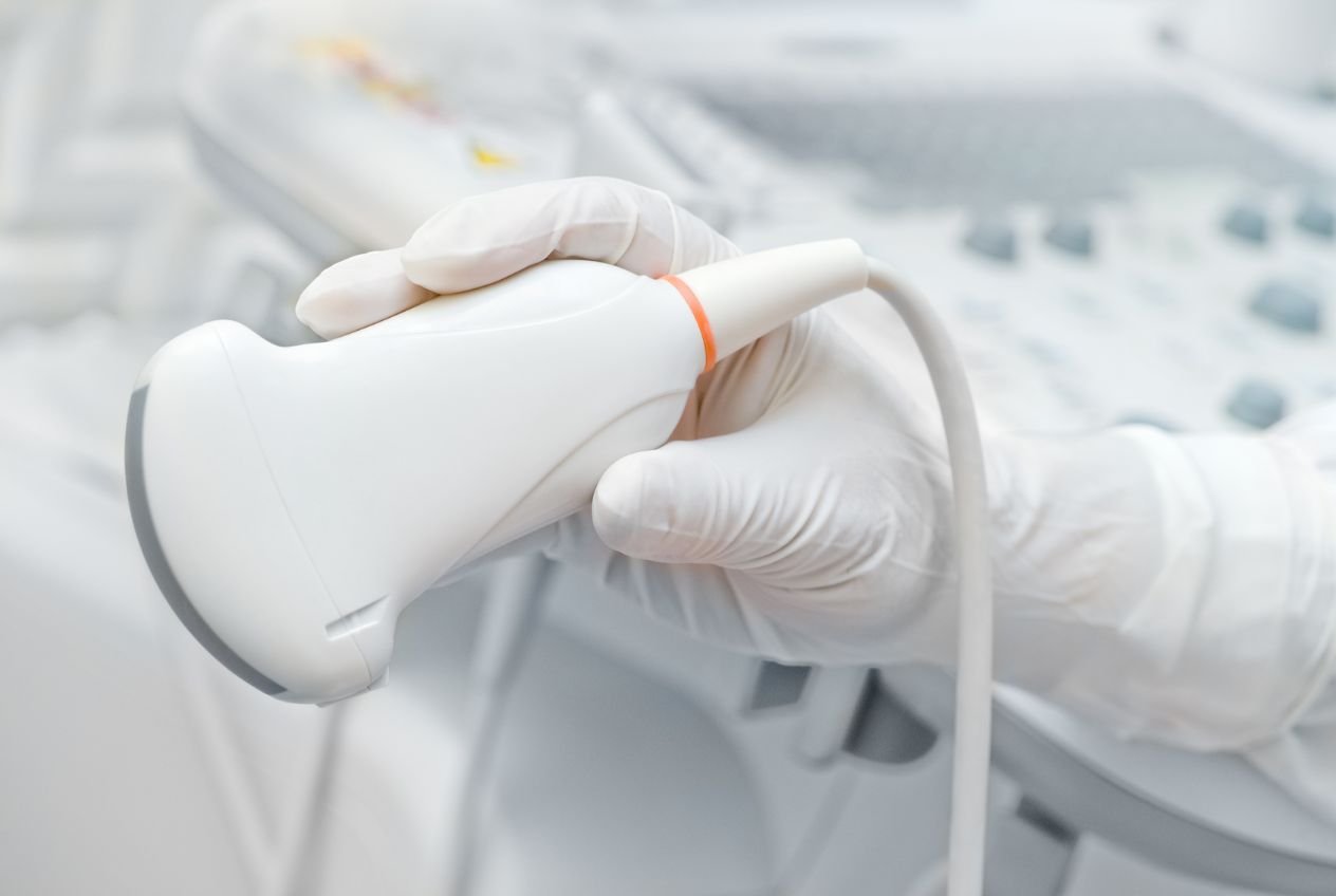
Ultrasound examination of the thyroid gland and soft tissues of the neck
Ultrasound examination with color doppler of the thyroid gland and soft tissues of the neck is a completely painless and non-invasive diagnostic method. Using high-frequency sound waves, this method gives us extremely precise data on the thyroid tissue and all surrounding structures.
In our polyclinic in Kragujevac, we are dedicated to detailed ultrasound diagnostics, which represent the gold standard and the absolute basis for early detection of all endocrinological disorders and changes in the neck region.
When should this examination be done? The examination is recommended for people who have a hereditary predisposition to thyroid diseases, as well as for the appearance of any visible enlargement, swelling or palpable nodules in the neck area.
Also, the examination is necessary in case of suspected inflammatory processes, as a regular control after operations in the neck area, but also in the case of other diseases that are closely related to the work of the thyroid gland (cardiac and hematological diseases, osteoporosis and unexplained variations in body weight).
What the examination reveals and the importance of color doppler
Using the ultrasound method, we get a clear image on the screen - a two-dimensional representation of the examined organs. On this occasion, the doctor not only observes the thyroid gland, but also the entire complex of soft tissues that surround it.
Structures to be inspected:
- Thyroid and parathyroid glands: Their size and shape are precisely measured and the homogeneity of the structure is assessed.
- Neck lymph nodes: Their size and appearance are examined in detail to rule out inflammation or malignancy.
- Salivary glands: Submaxillary and parotid glands.
- Muscles and sheaths: The condition of the connective tissue sheaths and neck muscles.
Importance of Color Doppler option:
This examination is always done on an ultrasound machine that also has the option of color doppler.
Thanks to this advanced technology, the doctor can measure the blood flow through the arteries that supply these organs. Obtaining a clear insight into the state of the blood supply of the examined organ or observed nodule is of key importance for distinguishing benign from potentially dangerous (malignant) changes.
What the examination looks like and the frequency of controls
The ultrasound examination is performed with the patient lying comfortably on his back, with his head slightly tilted back. The doctor gently passes the probe, on which a little ultrasound gel has been applied, over the skin of the neck. At the same time, the patient he does not feel any pain or discomfort. The examination itself lasts between 10 and 20 minutes, after which the patient receives an oral explanation and a detailed written report.
Like all other ultrasound methods used in modern diagnostics, this one is completely safe, there are no harmful effects (radiation) on the subject, which is why it can be repeated an unlimited number of times.
The time until the next check-up is determined by the doctor based solely on the findings. In the case of a normal finding, the next preventive control should be in a year, while in the case of a pathological finding (observed nodules or cysts), controls are scheduled for a month or several months for safe monitoring.